The voltage-gated sodium channel gene family
An introduction to the voltage-gated sodium channel gene family
Selective permeation of sodium ions (Na+) through voltage-dependent sodium (Nav) channels is fundamental for the generation of action potentials in excitable cells such as neurones and muscles. The voltage-gated sodium channel family is composed of nine pore-forming members, which are named Nav1.1 to Nav1.9 and encoded by genes referred to as SCN1A to SCN11A.
The voltage-gated sodium channel gene family
| Protein Name | Gene | Disease and therapeutic associations |
| Nav1.1 | SCN1A | Febrile epilepsy, Dravet syndrome, intractable childhood epilepsy with generalized tonic-clonic seizures, Panayiotopoulos syndrome, familial hemiplegic migraine, familial autism, Rasmussens's encephalitis and Lennox-Gastaut syndrome. |
| Nav1.2 | SCN2A | Inherited febrile seizures, epilepsy, and autism spectrum disorder. |
| Nav1.3 | SCN3A | Epilepsy and pain. |
| Nav1.4 | SCN4A | Hyperkalemic periodic paralysis, paramyotonia congenita, and potassium-aggravated myotonia. |
| Nav1.5 | SCN5A | Cardiac: Long QT syndrome, Brugada syndrome, and idiopathic ventricular fibrillation. Gastrointestinal: Irritable bowel syndrome. |
| Nav1.6 | SCN8A | Epilepsy. |
| Nav1.7 | SCN9A | Erythromelalgia, paroxysmal extreme pain disorder, channelopathy-associated insensitivity to pain and fibromyalgia. |
| Nav1.8 | SCN10A | Pain and neuropsychiatric disorders and small fibre neuropathy. |
| Nav1.9 | SCN11A | Pain |
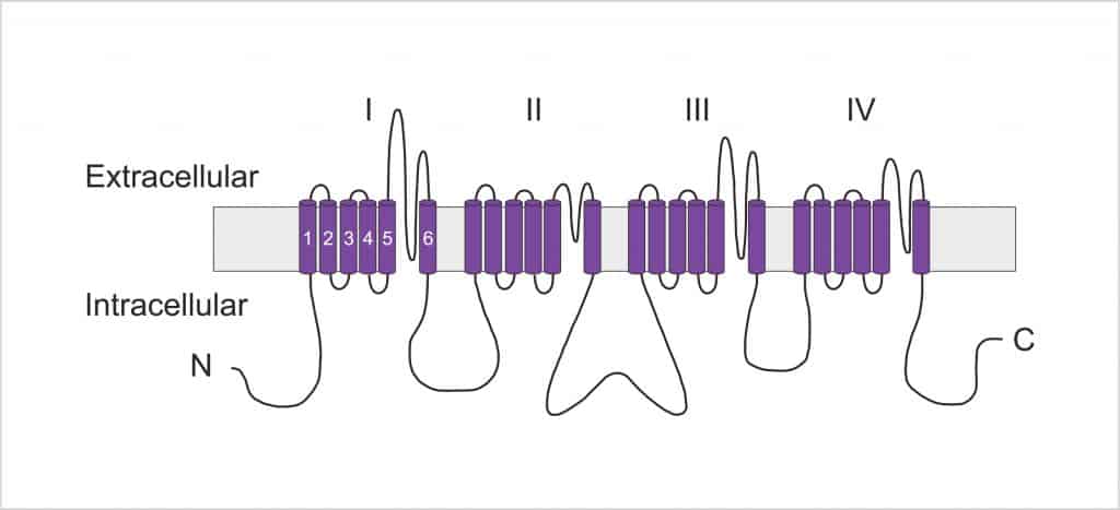
The channels consist of an α subunit – a large membrane spanning protein that forms the ion conduction pore – and one to two accessory (β) subunits. Whilst the α subunits are capable of forming functional channels, the association of β subunits can affect their biophysical properties (e.g. voltage-dependence) and cellular localization. The α-subunit is composed of four repeat domains, labelled I through IV, each containing six membrane-spanning segments, labelled S1 to S6. The highly conserved S4 segment acts as the channel’s voltage sensor whilst the S5-S6 loop helix incorporates the pore and the C-terminus inactivation gate.

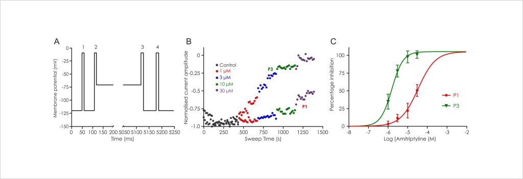
The important role of Nav channels in controlling the activity of excitable cells is supported by their involvement in various channelopathies (Table 1). This has fuelled a strong interest in the pharmaceutical industry to modulate the activity of sodium ion channels in a variety of therapeutic indications, especially pain and epilepsy. Fully understanding the electrophysiological properties of Nav channels is pivotal in developing efficacious and safe therapeutics. For example, companies focused on developing Nav channel inhibitors have tended to focus on identifying compounds that show a preference to the inactivated channel state, as Nav channels will accumulate in the inactivated state during periods of high frequency firing associated with patho-physiology. Metrion is highly experienced at developing custom Nav1.x screening assays to identify state-dependent inhibitors. Figure 2 shows representative data using a voltage protocol (Figure 2A) designed to evaluate the state-dependent inhibition of Nav channels. Four different concentrations of Amitriptyline were applied and the inhibition was determined for the first (P1; resting state inhibition) and the third (P3; inactivated state inhibition) depolarising steps (Figure 2B). Figure 2C shows the four-point concentration-response curves constructed using the inhibition data determined for currents elicited at P1 and P3. This voltage protocol demonstrated that Amitriptyline has a preference to the inactivated state of Nav channels. Metrion has similar assay validation data available for most Nav1.x channels (e.g. Nav1.7, Nav1.8 and Nav1.5), please enquire for more information.

Other recommended publications
Posters


Let’s work together
What are your specific ion channel and assay needs?
If you have any questions or would like to discuss your specific assay requirements, we will put you directly in touch with a member of our scientific team. Contact us today to discover more.
