Using Qube 384, we profiled a panel of NaV inhibitors across species, providing valuable translational insight early in analgesic drug discovery.
Metrion provides specialist neuroscience drug discovery services to biotech, pharmaceutical and academic organisations developing therapies for neurological disorders. Our expertise spans ion channel pharmacology, electrophysiology and translational neuroscience, supporting programmes from target validation through to lead optimisation and safety assessment.
Using a range of cellular, tissue and human iPSC-derived neuronal models, we help clients characterise compound activity, understand neuronal function and generate robust data to support drug discovery decision-making.
Our capabilities include native neuronal ion channel assays, neuronal firing assays, automated and manual patch-clamp electrophysiology, multi-electrode array (MEA) recordings and specialised translational neuroscience studies.
Pain research: Translational peripheral neuronal assays and platforms for pain research.
CNS drug discovery and brain slice electrophysiology: Increase the likelihood of successful therapeutic outcomes.
Native neuronal ion channel assays: Reliable and reproducible neuronal ion channel assays to demonstrate the efficacy, selectivity, and potency of therapeutic compounds.
Central neuronal firing assays: Reliable and reproducible translational neuroscience assays to demonstrate the efficacy, selectivity, potency and neurotoxic liability of your therapeutic compounds.
Translational neuroscience assays: Translational, phenotypic neuronal assays and platforms employing human iPSCs from the peripheral and central nervous system.
Cell line generation: Ready-to-go and customised cell lines engineered for reliable, reproducible screening success.


Our panel of models allow for more accurate prediction of human responses, reduce the risk of late-stage failures and provide a thorough understanding of drug mechanisms.

Figure 1. Electrophysiological recordings of primary neurons. A. Rodent dorsal root ganglion (DRG) neuron preparation for manual patch-clamp studies and a bright-field image of dissociated cells seeded on a coverslip. B. DRG action potential responses to low and high prolonged current injections.
A range of assay methodologies allows a greater understanding of the mechanism and impact of ion channel modulation on neuronal firing and network activity.
Examples of the assays we offer include:
Our neuroscience assays are used to:
Our neuroscience experts can evaluate potency and mechanism of action of compounds using heterologous cell lines expressing human or rodent neuronal ion channels (including challenging to develop cell reagents such as NaV1.8 and NaV1.9 lines). Effects of compounds on neuronal excitability can be assessed using human iPSC-derived neurons and primary rodent neurons.
Get insight into safety profiling of lead compounds and IND candidates, either as part of an existing screening cascade or because in vivo testing revealed a neurological signal. We have developed and validated an industry-standard rat cortical CNS neuron assay that can reliably detect the seizure-inducing liability of a wide range of reference compounds (as well as serve as a model to profile anti-epileptic drugs). We also have deep expertise with rodent dorsal root ganglion (DRG) sensory neurons which can be used to assess peripheral neurotoxicity.
Studying ion channels on the lysosomal membrane is essential for understanding lysosomal function, cellular homeostasis, and the development of various neurological diseases, such as Parkinson's disease. This research has the potential to uncover new therapeutic targets and improve our knowledge of fundamental cellular processes.
Read more about how we perform the lysosomal patch clamp technique to study ion channels on the lysosomal membrane, and watch this video to see it in action.
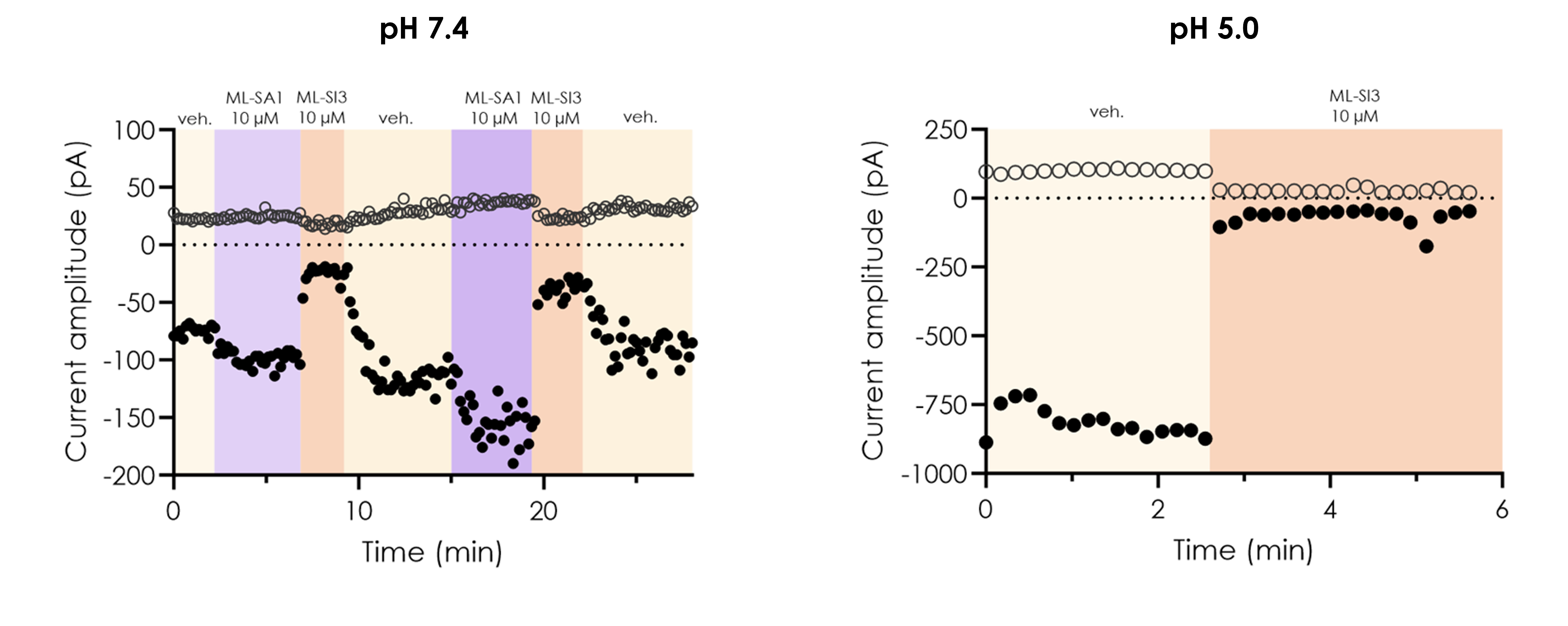
Figure 2. Pharmacological inhibition of wild-type TRPML1 currents recorded by manual patch-clamp.
Eliana is a two-year-old from Canada with a de novo mutation (V434L) in her KCNC1 gene which encodes for the Kv3.1 channel in central nervous system neurons such as cerebellar neurons and GABAergic interneurons. The mutation manifests as a variety of neurological disorders which can include myoclonic epilepsy and ataxia, developmental epileptic encephalopathy (DEE), or hypotonia, depending on the specific variant.
The KCNC1 Foundation collaborated with Perlara, who approached Metrion Biosciences, where manual and automated (Qube) patch-clamp techniques and Fluorescent Imaging Plate Reader (FLIPR) high-throughput screens (HTS) against the mutant channel were performed to identify hit compounds.
Metrion provides neuroscience drug discovery services including pain research, CNS drug discovery and brain slice electrophysiology, native neuronal ion channel assays, neuronal firing, translational assays.
Our studies utilise human iPSC-derived neurons, primary cortical neurons, trigeminal neurons, dorsal root ganglion neurons, recombinant ion channel cell lines and brain slice preparations.
Translational neuroscience assays evaluate compound effects in physiologically relevant neuronal systems to help predict clinical outcomes and bridge the gap between target-based screening and in vivo studies.
Ion channels can be studied using manual patch-clamp electrophysiology, automated patch-clamp platforms, neuronal firing assays and multi-electrode array recordings.
Pain drug discovery programmes commonly utilise DRG neuron electrophysiology, peripheral neuronal firing assays, NaV channel screening, automated patch-clamp studies and translational sensory neuron models.
Using Qube 384, we profiled a panel of NaV inhibitors across species, providing valuable translational insight early in analgesic drug discovery.
We explore hNav1.9's unique fast and slow inactivation properties using Qube 384 and QPatch 48 platforms, helping to build more predictive screening assays for state-dependent inhibitors.