GLP hERG screening assay validation following ICH E14/S7B 2022 Q&A best practice guidelines

Case Study
Case Study title
GLP hERG screening assay validation following ICH E14/S7B 2022 Q&A best practice guidelines
Authors
James Masterson, Sergiy Tokar, Ayesha Jinat, Robert Kirby
Metrion Biosciences Ltd, Granta Centre, Granta Park, Cambridge, CB21 6AL, United Kingdom
Overview
The ICH E14/S7B 2022 Q&As provide the best practice guidelines for evaluating the effect of preclinical compounds on the human ether-à-go-go-related gene (hERG) potassium channel1. The guidelines stipulate that in vitro hERG assessments should be performed to Good Laboratory Practice (GLP) compliance. In addition, they provide recommendations on the experimental methods that should be employed, the quality control parameters for analysing the data, as well as the preferred format for reporting the data. This is to ensure data quality, transparency and consistency throughout the industry.
A hERG assay is considered negative if the safety margin calculated for the test article is greater than the established safety margins generated with the Food and Drug Administration’s (FDA’s) positive controls (ondansetron, moxifloxacin and dofetilide) tested to the best practice guidelines. Metrion conducted a GLP compliant study using the conventional manual patch-clamp technique in accordance with the ICH E14/S7B Q&A best practice guidelines to establish in-house IC50 values for ondansetron, moxifloxacin and dofetilide.
Materials and methods
Cell preparation
- Chinese hamster ovary (CHO) cells stably expressing the hERG1a isoform were plated onto glass coverslips and cultured at 37 °C for 24 – 48 hours before being used for electrophysiological recordings.
Recording solutions
- Intracellular (in mM): 120 K-gluconate, 20 KCl, 10 HEPES, 5 EGTA, 5 MgATP; pH 7.3
- Extracellular (in mM): 130 NaCl, 10 HEPES, 5 KCl, 1 MgCl2*6H2O, 1 CaCl2*2H2O, 12.5 dextrose; pH 7.4
Compound preparation
- Moxifloxacin was dissolved in extracellular solution and stock solutions were stored at -20 °C. A new aliquot was thawed each experimental day and diluted to generate each test concentration.
- Ondansetron and dofetilide were dissolved in 100% DMSO and stored at -20 °C. A new aliquot was thawed each experimental day and diluted in extracellular solution (0.1% DMSO final concentration) to generate each test concentration.
Electrophysiology
- Whole-cell voltage-clamp experiments were performed at 36 ± 1 °C using a HEKA EPC10 patch- clamp amplifier and PatchMaster Pro (version V2.73) software (HEKA Elektronik).
- Voltage errors were minimized by using ≥ 80% series resistance (Rs) compensation.
- Membrane currents were sampled at 20 kHz and filtered using double stage low pass Bessel filters set at 10 kHz and 2.9 kHz .
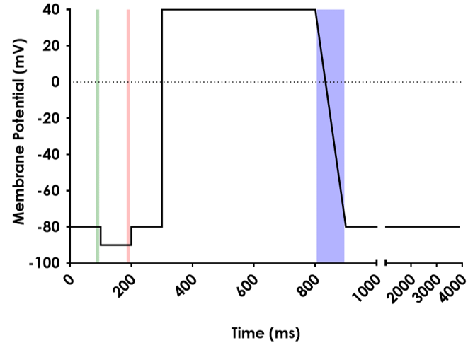
- Membrane currents were elicited using the “Recommended voltage protocol to study drug-cardiac ion channel interactions using recombinant cell lines” FDA 2021 (Figure 1), which was applied at
- 0.2 Hz. The voltage command was adjusted for the liquid junction potential (-15 mV).
Figure 1. The voltage protocol that was used to elicit hERG current. The shaded areas show the location of the cursors used to analyse:
Green: Holding current
Red: Current elicited by step to -90mV
Blue: Peak current.

Compound application
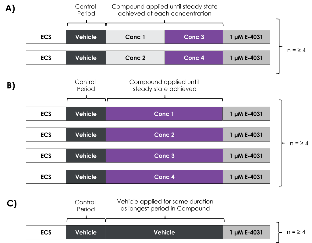
- After obtaining the whole-cell configuration, the cells were superfused with extracellular solution containing vehicle (0.1% DMSO) to allow the current to stabilise (Control Period).
- Following the stabilisation period, the compounds were evaluated using a two-point assay format (Fig 2A; ondansetron and moxifloxacin) or a single-point assay format (Fig 2B; dofetilide).
- Each experiment was completed with the application of 1 µM E-4031, which allowed the off-line subtraction of leak and endogenous current.
- Vehicle controls, matched to the longest recording in compound, were also performed (Fig 2C).

Quality control
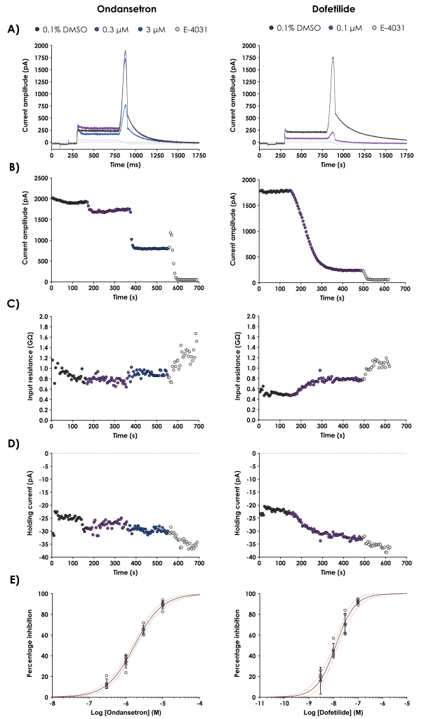
- The peak current amplitude, input resistance and holding current were plotted against time and reviewed for each cell. Cells with large changes in these parameters were discarded.
- In addition, the Peak Current amplitude and stability during the Control Period, series resistance, bath temperature and the inhibition produced by 1 µM E-4031 were also used to QC the data.
- Samples of the compounds were collected after each experiment, which allowed concentration verification to be performed for each concentration of the compounds.
Data analysis
- Percentage inhibition of the peak current amplitude was calculated using E-4031 subtracted current values.
- The mean (± SD) inhibition data were plotted against concentration (on a logarithmic scale) and fitted with the following four-parameter logistic fit equation (A = 0, B = 100, C = pIC50, D = Hill slope):

- In addition, 95% confidence bands were included on each graph, as well as the individual inhibition values calculated for each concentration of the compounds.
Results
Figure 3: Representative current traces.

| Ondansetron | Moxifloxacin | Dofetilide | Vehicle | |
| Peak Current (pA) | 1535.3 ± 242.5 | 1257.9 ± 119.1 | 1360.2 ± 170.9 | 1324.1 ± 242.4 |
| Input Resistance (MΩ) | 770 ± 90 | 580 ± 30 | 560 ± 50 | 680 ± 80 |
| Series Resistance (MΩ) | 3.5 ± 2.1 | 4.0 ± 3.0 | 3.2 ± 2.1 | 3.2 ± 1.3 |
| Holding Current (pA) | -32.1 ± 5.5 | -37.1 ± 6.4 | -25.5 ± 3.6 | -18.4 ± 6.9 |
| Compound | ICH E14/S7B training material IC50 (µM) (95% confidence intervals) | Metrion IC50 (µM) (95% confidence intervals) |
| Moxifloxacin | 62 (38, 104) | 96.2 (78.6, 117.7 ) |
| Ondansetron | 1.4 (0.8, 2.6) | 1.72 (1.51, 1.95) |
| Dofetilide | 0.01 (<0.01, 0.02) | 0.012 (0.011, 0.013) |
Summary
- Ondansetron, moxifloxacin and dofetilide were successfully validated using the ICH E14/S7B Q&A best practice guidelines and to UK GLP compliance.
- The IC50 values generated in this study were within 2-fold of the values reported in the ICH E14/S7B training material. This result allows Metrion to use the FDA’s reported pooled reference drug safety margin of 51-fold higher than the free Cmax for test articles.
- In another (non-GLP) study performed to the ICH E14/S7B Q&A best practice guidelines, Metrion obtained an IC50 for ondansetron of 1.55 µM, which demonstrates the reproducibility of the assay.


Let’s work together
What are your specific ion channel and assay needs?
If you have any questions or would like to discuss your specific assay requirements, we will put you directly in touch with a member of our scientific team. Contact us today to discover more.
