Review written by the Editor
ELRIG is a not-for-profit organisation aiming to provide access to high level scientific content, promote innovation in drug discovery and provide networking platforms for the life science community. To implement this, ELRIG hosts conferences throughout the year across various sites, bringing together life science focused researchers from across Europe.

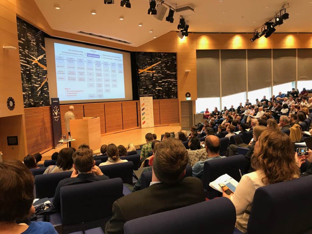
‘Advances in Cell Based Screening’, hosted at AstraZeneca’s impressive Mölndal based facility is one of five 2019 ELRIG hosted conferences. This event was focused heavily on assay precision, a topic which spans screening, target validation and drug development. Multiplexed and phenotypic screening were particularly highlighted within several of the presentations, as were novel techniques such as Cell Painting.
Whilst many of the presentations were not within Metrion’s key focus area of ion channel drug discovery, it was an excellent opportunity to hear about emerging new techniques and the breadth of opportunities to create assays with highly precise readouts for a range of druggable targets. I will summarise below a selection of the talks including three of the keynote presentations.
Target Engagement and Coherence with Functional Cellular Responses
The keynote on Day one within the session entitled ‘Target Engagement and Coherence with Functional Cellular Responses’ was given by Professor Herbert Waldmann of the Max Planck Institute of Molecular Physiology. He spoke on the topic of ‘Chemotype, Phenotype, Target’. The lab lead by Professor Waldmann are focused in part on the development of natural product (NP) inspired compound collections using Biology Oriented Synthesis (BiOS).
He discussed the utility of assays focused around the conversion of phenotypes and their relevance in locating unbiased novel targets. He confirmed the relevance of natural products in drug discovery, explaining that in their 2014 publication (Nat. Rev. Drug. Disc. 2014, 13, 577), Eder, Sedrani and Wiesmann from Novartis had analysed the origins of drugs approved by the FDA between 1999 and 2013 and found that 30% are biologics, 13% are natural products and 15% are natural substance derived molecules.
Hedgehog pathway inhibitors
Professor Waldmann discussed his work on hedgehog pathway inhibitors. The group created an osteoblast differentiation assay using purmorphamine (the first small-molecule agonist developed for the protein Smoothened (Smo), a key part of the hedgehog signalling pathway) as pathway agonist. Using C3H cells they generated a hedgehog pathway assay and observed Smo binding using Vismodegib (used to treat basal cell carcinoma) stained with BODIPY dye. To find the target they used quantitative proteomics (label free) to look for differences between the inactive and active probe.
The Waldmann lab have also been using cell painting assays (used to monitor changes in cellular features) to stain cellular compartments, generating five different fluorescent channels. Images are then analysed using a cell profiler. They found that many compounds are able to enter the lysosome, as they are protonated and trapped. In general, cell painting is a useful tool due to its ability to identify the phenotypic impact of chemical/genetic perturbations, collecting compounds/genes into functional pathways and identifying disease signatures.
Imaging single cell target engagement
Later within this track, Dr Matt Dubach, from Harvard University spoke about imaging single cell target engagement. He discussed a recent project focused on an in vivo approach to the study of pharmacokinetics and target engagement. The group are specialists at cellular level drug imaging and presented an example in which intravital microscopy can be used to image the PARP inhibitor Olaparib tagged with BODIPY to gain information on drug distribution.
The group use fluorescent microscopy and plate-based assays to further elucidate drug mechanisms and Dr Dubach displayed images of fluorescent drug being added to cells and how nuclear staining can be observed. By calculating the anisotropy, the group were able to measure drug binding. They progressed their study to the use of clinical Olaparib and using a Schild analysis, they could determine KDs accurate with in vitro protein based measurements. This helps to elucidate drug binding mechanisms within cells and helps to deduce the optimal drug type for tumour treatment.
The final step of the project was to image in vivo and they performed controlled delivery to administer clinical and fluorescent drug to tumours. Results indicated that fluorescent drug was essentially competed off by the clinical drug. Dr Dubach discussed their in vivo versus in vitro studies. They saw an increased heterogeneity in vivo and concluded that the experiment is affected by expression levels and drug distribution which is not uniform. In this type of study, to generate the most reliable data, the clinical drug should be injected at different doses and fractional occupancy measured.
Bulk measurements can be misleading, as they can indicate saturation of the target. However, at the single cell level, experiments showed that cells encounter some protein inhibition, but still possess enough non-inhibited protein to permit function (for example tumour cells can still grow), known as fractional occupancy. With their approach one can look at single cell heterogeneity, this works with non-covalent drugs in vivo, which enables other quantitative measurements.
Precision Medicine in Miniaturized Format
During the afternoon track ‘Precision Medicine in Miniaturized Format’, the keynote presentation was given by Professor Olli Kallioniemi from SciLifeLab and the Karolinska Institute who spoke on the topic of ‘Functional Precision Medicine for Cancer and Beyond’. He explained that disease characteristics are changing as a result of molecular information and hence we can sub-classify diseases to make better treatment decisions.
Prof. Kallioniemi elaborated on a pilot study based around the concept of 14 dimensions of health which looks at 14 correlated molecular features which may contribute to the overall health of individuals. Factors such as diet, the gut microbiome, alcohol consumption and blood pressure are considered with the aim of combining this information, drawn from various diverse sources and considering complex inter-connections of genome, proteome, microbiome, metabolome, diet and lifestyle. People who appeared to be in poor health were offered advice to improve certain characteristics where possible.
Professor Kallioniemi next described the challenges of predicting cancer drug responses in patients. 10-15% of all cancers could be helped by having a druggable clinical mutation, but of course many of the common mutations such as P53 are not ‘directly druggable’. He elaborated on functional precision systems medicine in leukaemia and emphasised the need to better understand the biology of the disease, drug sensitivity and resistance, how to rapidly induce therapies and then to report data back to the clinician.
He described a study in which they took 500 known cancer drugs, performed dose response testing in patient derived cells (plate reader based), looking at viability, toxicity and other characteristics and then compared drug efficacy on normal cells using bone marrow. This is known as ‘ex vivo drug testing’. It allows the user to gain results on the comparative efficacy, patient profile and biomarkers for each drug and to formulate drug efficacy correlations. One can also learn about mechanism of action, resistance and the evolution of the drug response over time. This can be run alongside clinical trials and would also allow the user to identify new effective drugs if resistance has occurred.
Accelerating Drug Discovery through the power of microscopy images
Day Two’s keynote talk was presented by Dr Shantanu Singh of The Broad Institute and was entitled ‘Accelerating Drug Discovery through the power of microscopy images’. Dr Singh described the various imaging techniques used at the Broad to measure and score known phenotypes, profile and characterise samples and analyse the data generated. They can then identify compounds to use in treatments. He described a cell painting assay they developed using six stains, imaging five channels and revealing eight constituents/ organelles.
By using an automated microscope, they can also confirm signatures of genes, compounds and diseases. Dr Singh elaborated on how image-based profiling can predict small molecule activity. He described a 1000 compound screen carried out using cell painting techniques, with the resulting images then being matched to morphological profiles. This project is still underway at the Broad.
Dr Singh explained that the Broad are also focused on rare diseases. Of 7,000 known rare diseases, 4,550 are known to be monogenic and only 6% have FDA approved treatments. He discussed whether it would be possible to screen a vast number of compounds using cell painting techniques, create a database and determine which give the disease signature. This is known as image-based profiling for virtual screens.
Bioimaging is poised for dramatic improvements driven by deep learning. This includes deep learning for the segmentation of nuclei. In collaboration with Peter Horvath’s lab, The Singh lab created a new deep learning tool known as Cyto AI. The lab have also been progressing another project focused on the extraction of features by training networks on auxiliary tasks. This allows compound grouping based on their mechanisms. They are also able to capture the heterogeneity accurately by modelling data statistics and using a wet lab method known as pooled cell painting, can scale up profiling of genes via in situ sequencing.
Advances in Phenotypic screening
Later in the session, Professor Neil Carragher from the University of Edinburgh described ‘Advances in Phenotypic screening: Accelerating the Discovery of New Chemical Entities and Drug Combinations towards in vivo proof of concept’. Professor Carragher explained that the Proteomics Drug Discovery group is predominantly involved in cell and tissue-based screening assays to validate novel targets.
The lab used the image Xpress (PAA robotic), with cell profiler analysis to analyse tens of thousands of small molecules, in order to create phenotypic fingerprints for every compound and then complete machine learning to predict the mechanism of action. He described how they trained the machine learning models in breast cancer cell lines and applied the model to unseen cell lines and discussed a method for comparing the similarity of compounds to differential cellular phenotypic responses across breast cancer cell lines.
Professor Carragher then described a case study collaboration with CRUK (Rebecca Fitzgerald, Ted Hupp, Rob O’Neill) for a drug discovery project on oesophageal cancer. He explained that they undertook a high throughput screen of 20,000 chemical libraries, using a cell line panel that represents the heterogeneity of disease. Using a machine learning model combined with the feature extraction method, they saw novel phenotypic space and mode of action. Using morphometric profiles, they were able to detect compounds with selectivity for oesophageal cells.
The conference was interspersed with networking sessions and delegates also enjoyed a drinks reception on the first evening. During these more informal breaks, scientists were able to discuss their research and their thoughts on the talks, meet with vendors and make valuable connections in addition to catching up with existing contacts. A tour of AstraZeneca’s facility was offered on the evening of Day two and Day three followed the format of a satellite meeting which was focused around Chemical Biology, namely PROTACs as a novel therapeutic modality and proteomic target identification strategies.
Metrion Biosciences now looks forward to exhibiting and presenting at the largest ELRIG event of the year, ‘Drug Discovery: Looking back to the Future’ to be held at the ACC in Liverpool on 5th and 6th November.
